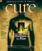
Esophageal Cancer: A Disease on the Rise
Fighting esophageal cancer starts by addressing the reasons behind it.
Esophageal cancer is an aggressive form of cancer, and one that often remains asymptomatic until relatively late in the disease process. “Before I was diagnosed with this, I had never heard of it,” says Bart Frazzitta, from Manalapan, New Jersey. “I didn’t even think you could get esophageal cancer.” In fact, there were approximately 14,250 new cases of esophageal cancer diagnosed in 2004 in the United States and 13,300 deaths from the disease. Additionally, of all the solid tumors, esophageal cancer is the one that has been increasing most rapidly in recent years. The reasons for the increase may be related to an increase in gastroesophageal reflux disease and other lifestyle factors.
Esophageal cancer initially develops in the esophagus, which is the smooth muscular tube that carries food and liquids from the throat to the stomach. There are two types of esophageal cancer: squamous cell carcinoma and adenocarcinoma. Squamous cell carcinoma occurs when the cells that normally line the inside of the esophagus begin to proliferate abnormally. Adenocarcinoma develops from abnormal glandular cells that line the walls of the esophagus that has been damaged by gastroesophageal reflux disease (GERD).
Previously, squamous cell carcinoma was the predominant form of the disease. In recent decades, however, adenocarcinoma has become more prevalent and now accounts for more than half of all cases of esophageal cancer in the United States, says Stuart Spechler, MD, from the University of Texas Southwestern Medical Center in Dallas.
Unfortunately, the disease does not produce many symptoms in its early stages and thus goes undetected. Large, advanced tumors may cause weight loss, difficulty swallowing, or evidence of blood in stool or vomit.
With no early warning symptoms, individuals often don’t realize something serious is going on until they have advanced cancer. Frazzitta’s only symptom was chronic heartburn a couple of times a week for about a year. Yet when he was diagnosed in December 1999, he had stage 3 cancer, which means the disease had already spread from the esophagus to surrounding tissues or lymph nodes.
“A lot of people will go and get an antacid and feel better and think they have cured the problem,” says Frazzitta, who subsequently has become a patient advocate and co-founder of the Esophageal Cancer Education Foundation (www.fightec.org). In truth, the antacid just masked a symptom of a serious disease, which is why experts recommend that if heartburn or indigestion persists, people should see a doctor.
The two types of esophageal cancer occur with different frequencies in different ethnic groups. Squamous cell carcinoma is more common in blacks and Asians, while white men are at highest risk for adenocarcinoma.
Some of the risk factors for the two subtypes are the same, but not all of them. For example, obesity is a risk factor for adenocarcinoma, while malnutrition puts individuals at increased risk for squamous cell carcinoma. However, heavy smoking and age over 50 are associated with an increased risk of both types of disease.
Sometimes, however, even those risk factors don’t apply. Vickie Powell, a resident of Radcliff, Kentucky, was just 38 when she developed a case of the hiccups that she couldn’t keep at bay. Initially her doctor put her on an antacid, which she took for a week and a half.
"You know your own body," says Powell. “I went back to the doctor and said something just is not right.” After more tests, she was told that she had a golf ball-sized tumor in her lower esophagus and upper stomach. She wasn’t overweight, she didn’t drink or smoke, and she doesn’t ever remember having heartburn.
A major risk factor for adenocarcinoma of the esophagus is Barrett’s esophagus, which occurs when the valve that lies between the stomach and the esophagus doesn’t function properly. Normally, it acts like a one-way gate, letting food and liquid move from the esophagus into the stomach, but keeping the acid and digestive enzymes of the stomach in the stomach. If the valve weakens, then acid will escape into the esophagus, which is called acid reflux. Repeated exposure to the acid damages the esophageal cells, causing them to die.
When new cells arise to take their place, a strange thing happens, says Dr. Spechler, who is an expert on Barrett’s esophagus. Instead of regenerating new esophageal cells that would continue to be damaged by the acid escaping from the stomach, the esophagus begins to produce cells that resemble those that line the intestinal tract. Such cells are ready for an acid bath and aren’t damaged as easily by acid reflux.
Unfortunately, these cells are predisposed to form cancers when they arise in the esophagus. Thus, patients with chronic GERD and who develop Barrett’s esophagus are at significantly higher risk for esophageal cancer and should undergo regular cancer surveillance. One common form of surveillance is called endoscopy, in which a physician runs a thin tube with a camera and a light at the end into the esophagus, allowing for a visual of what the tissue looks like.
Screening all GERD patients with endoscopy to look for Barrett’s esophagus is neither feasible nor practical, however. Approximately 60 million adults in the United States have regular heartburn, but only a tiny fraction of those will develop esophageal cancer. Researchers are trying to identify better ways to diagnose esophageal cancer earlier.
Like many cancers, the timing of diagnosis is critical in esophageal cancer. “If we can catch it early, we can cure it,” says Dr. Spechler. With that in mind, physicians like to closely follow patients who have Barrett’s esophagus or other high-risk factors for the disease.
If the cells in the esophagus begin to look abnormal but are not yet cancerous, a stage called dysplasia, Dr. Spechler and others will often recommend surgical treatment to prevent full-blown cancer from developing. If left untreated, 31 to 59 percent of patients with high-grade dysplasia will develop cancer within five years.
Currently, the standard surgery for high-grade dysplasia is an esophagectomy, in which the surgeon removes the unhealthy portion of the esophagus and possibly some of the neighboring stomach, and then reconnects the remaining regions of the tube together. Although this surgery is effective at preventing the disease and treating some stages of the cancer itself, it is a difficult surgery and between 3 and 12 percent of patients may die as a result of the surgery itself.
Researchers are testing less drastic surgical methods, but these are still in clinical trials, and it is not yet clear how effective they are at preventing the development of cancer, although early results look promising. One method involves using an endoscope with a small blade attached to the end to cut away the unhealthy cells. Another method being tested is to use a laser to burn away the dysplastic cells.
For those patients who are diagnosed with esophageal cancer, esophagectomy is the standard therapy, unless the cancer has spread beyond the esophagus and adjacent tissue. The proportion of esophageal cancer patients alive five years after diagnosis is between 5 and 30 percent, depending on the stage of cancer they have at diagnosis and how well they respond to treatment.
Most patients with localized esophageal cancer (stage 1 or 2 disease) used to be treated with surgery alone. In recent years, a combination of chemotherapy and radiation or chemotherapy and radiation followed by surgery is being used more often to treat esophageal cancer, says David Ilson, MD, from Memorial Sloan-Kettering Cancer Center in New York. The addition of chemotherapy to a radiation treatment protocol can enhance the effectiveness of radiation and increase the proportion of patients who achieve a complete remission or a good partial remission with radiation. Subsequent surgery, done after chemotherapy and radiation, can then be used to remove any residual cancer. Combining chemotherapy, radiation therapy and surgery (called combined modality approaches) are increasing the cure rate of this cancer.
The current standard chemotherapy given with radiation therapy is a combination of 5-FU plus cisplatin, but this can lead to significant side effects of mouth sores and esophagitis. “We are trying to identify more effective chemotherapy regimens that have fewer side effects,” says Dr. Ilson.
Regimens currently being tested use cisplatin in combination with newer chemotherapy drugs like Camptosar® (irinotecan), Taxotere® (docetaxel), Taxol® (paclitaxel) and Gemzar® (gemcitabine). Since cisplatin causes a number of side effects, including nausea, vomiting, kidney damage and hearing loss, a better-tolerated analogue of platinum oxaliplatin (Eloxatin™) is being studied in esophageal cancer.
For patients with metastatic (stage 4) disease, surgery is not recommended. Rather, physicians treat such advanced disease with chemotherapy, which is not likely to cure the cancer but may slow its progress and make the patient more comfortable.
Chemotherapy drugs that are used include 5-FU, cisplatin, Taxol, Taxotere, Camptosar and Eloxatin. Additionally, a phase III randomized trial in the United Kingdom is testing the efficacy of a Xeloda® (capecitabine) combination in esophageal cancer. But Dr. Ilson says Xeloda is not yet used frequently in the United States for this disease.
In addition to traditional chemotherapy agents, physicians are testing the use of newer, less toxic targeted drugs in esophageal cancer. Unlike standard chemotherapy drugs, which kill all dividing cells, targeted therapies block specific steps in the cancer cell growth pathway and leave most healthy cells undamaged.
Dr. Ilson’s research group is setting up trials to test the monoclonal antibody Erbitux™ (cetuximab), which targets the epidermal growth factor receptor (EGFR), in combination with standard chemotherapy in patients with locally advanced esophageal cancer. They will also test Avastin® (bevacizumab), another monoclonal antibody that blocks blood vessel formation, in combination with chemotherapy and radiation in adenocarcinoma-type esophageal cancer.
Patients with advanced esophageal cancer may experience difficulty swallowing and an inability to take solid food, which can cause malnourishment and an inability to tolerate chemotherapy. In patients who are not candidates for curative surgery or radiation-based treatment, the relief of swallowing difficulties is a primary goal of palliative or symptom-relieving management. Chemotherapy alone in advanced disease may relieve swallowing problems.
Additionally, there are several options for local therapy, and esophageal stenting is one of the most common. During the procedure, a physician uses endoscopy to place a stent in the esophagus, and, because the stent is a semi-rigid tube, it prevents the tumor from blocking or narrowing the esophagus.
Another option that is sometimes used is called photodynamic therapy. In photodynamic therapy, the patient receives an injection of a drug called Photofrin® (porfimer sodium) that causes cells to be sensitive to light. The gastroenterologist uses an endoscope to then shine laser light on the tumor, killing the cancer cells. One problem with this approach is that the patient has to avoid light for several weeks after treatment, which can significantly lower his or her quality of life. Also, the results of this treatment are relatively short-lived.
If a patient has failed chemotherapy or they have a stent in place but the tumor is overgrowing the stent, physicians may use radiation to locally control the tumor. In some cases, the doctor may opt to use laser therapy in which a laser is attached to an endoscope to kill off parts of the tumor that are causing swallowing difficulties. In this situation, however, radiation is used more frequently than lasers, says Dr. Ilson.
For patients who are malnourished and not able to take in adequate amounts of calories, a feeding tube is placed in the stomach. This tube (usually called a PEG tube) allows food to be administered directly into the stomach, thus ensuring the patient receives adequate nutrition.
Until less toxic treatments are available, the emotional and physical cost of therapy can be high. For example, Powell had an esophagogastrectomy in which the surgeons removed one-fifth of her stomach, 8 inches of her esophagus, the surrounding lymph nodes and the sphincter muscle that controls flow between the esophagus and the stomach. Additionally, because she was so young, her doctor wanted to use a particularly aggressive therapy to prevent recurrence. Thus, after surgery, she received two rounds of chemotherapy, with each round consisting of five days of continuous intravenous drip of 5-FU and cisplatin. She also had 30 radiation treatments.
Now, as she has just had her seventh-year CT scan, she remains cancer-free, though she suffers from surgery complications. Without a sphincter muscle, she still has trouble keeping food down and has to eat small meals every couple of hours rather than three normal-sized ones. And even the small ones make her nauseous. “For a while I was really depressed about it, but I have learned to deal with it. This is what I have left. But I am cancer-free and I try to move on every day,” she says. “Sometimes I don’t realize how lucky I am until I go to the doctor and I tell my story. Now, I’m just at a point where I want to help as many people as I can.”
Frazzitta, who participants can meet at the CURE Patient & Survivor Forum in Washington, D.C., this July, had six weeks of Taxol and cisplatin chemotherapy plus radiation to treat his stage 3 disease. After recovering from this therapy for two months, he underwent an esophagogastrectomy in which the surgeon removed two-thirds of his esophagus and one-third of his stomach. He left the hospital 10 days later.
That was May 2000. Now, nearly five years later he won’t say he’s cancer-free, but his last CT scan six months ago was clear—and he hopes the next one will be clear as well.