Publication
Article
CURE
Cancer Imaging Gets Sophisticated
Author(s):
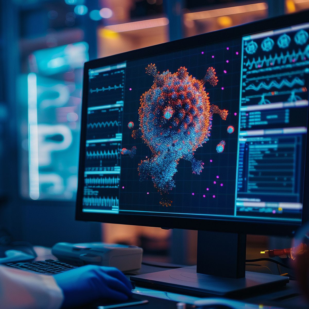
The next generation of cancer imaging
While today’s imaging tests provide anatomical information about a tumor’s size and location, tomorrow’s imaging technologies will look deeper, at the molecular make-up of cancer cells.
Some potential uses for these more advanced technologies include detecting and characterizing cancers without the need for biopsies or other invasive procedures, ensuring that drugs are reaching the tumor and promptly monitoring the effect of therapies so treatment can be changed if necessary.
Positron emission tomography (PET) is leading the way in the molecular imaging revolution. Currently, in oncology most PET imaging is performed with a radioactively-labeled form of glucose, a type of sugar, called 18F-fluorodeoxyglucose, or simply FDG. Cancer cells, particularly those from aggressive tumors, metabolize significantly larger amounts of glucose than surrounding normal tissue. FDG-PET imaging measures glucose uptake, revealing how active a cancer is and detecting whether the cancer has spread beyond its original location.
Glucose metabolism is not unique to cancer cells; however, and not all cancers exhibit high metabolic rates, so researchers are working on developing agents that are much more specific. For example, scientists are studying how to detect when cells are dividing quickly or when they are dying as a means of determining whether a cancer treatment is effective. Others are looking at ways to target proteins on the surface of cancer cells, such as the HER2 receptor, the epidermal growth factor receptor (EGFR) or the estrogen and androgen receptors. Finding out whether the expression of these receptors increase or decrease when a cancer spreads may eliminate the need for repeated biopsies and allow therapies to be tailored to the individual cancer at each stage.
New agents are also being developed to visualize angiogenesis, or growth of blood vessels within and around tumors. This research may be useful in monitoring whether antiangiogenic therapies are working, or identifying which patients might best benefit from such therapy.
According to David M. Schuster, MD, director of nuclear medicine and molecular imaging at Emory University Hospital, “all of these agents are just experimental right now and studies are being done, but I think these new agents, especially coupled with advanced devices, may hold promise in the future — maybe not for mass screening, but in problem-solving situations and in helping to individualize therapy.”
Also on the horizon: molecularly-targeted dyes and probes that can work with common optical imaging systems. Fluorescent dyes that attach to proteins found on cancer cells are being tested with specially modified microscopes for use in screening procedures, such as colonoscopy or endoscopy. These dyes may be able to determine more quickly if an abnormal growth is cancerous.
In one early test, a colon cancer-specific dye showed a high degree of sensitivity and specificity for detecting cancerous cells in the colon. Another promising light-based technique uses a light source to create a high resolution, cross-sectional or three-dimensional image that can “see” 1 to 2 millimeters below the surface. The technique, called optical coherence tomography (OCT), is being investigated for the identification and surveillance of Barrett’s esophagus. Imaging tests like these could also be used for other cancers that can be reached with endoscopes or catheters, such as esophageal or bladder cancers.
Ultrasound and magnetic resonance imaging (MRI) tests are also getting upgrades. Non-specific contrast agents are currently used in contrast-enhanced endoscopic ultrasound to increase the ability of ultrasound to visualize tumors with low blood flow, such as pancreatic cancers. Now, next-generation molecularly targeted “microbubbles” are being designed to visualize disease-specific molecules in blood vessels and could be used to monitor response to antiangiogenic therapies. Likewise, molecularly-targeted magnetic particles are being developed for use with MRI.
“I think the most exciting area, though, is hybrid technology, where we take anatomic imaging techniques, such as MRI, and bring them together with molecular techniques,” Schuster says. “For example, combine mammography, say tomosynthesis, with using radiotracers—and these are all other areas, which also have great promise.”
Other hybrid techniques that are being tested include PET-MRI, which was recently cleared for use in June by the Food and Drug Administration, single-photon emission computed tomography (SPECT)-CT and MRI combined with optical fluorescence technology.
Although most of these techniques are still experimental, researchers are hopeful that advances in imaging detection systems will further improve the lives of patients with cancer through earlier detection, when the cancer is at a more curable stage, as well as better management and personalization of treatment for cancers at a more advanced stage.