|Articles|May 1, 2017
Non-Invasive Imaging Device Brings a New Dimension to Early Skin Cancer Detection
Author(s)Katie Kosko
Non-invasive imaging devices can help dermatologists examine the skin and a lesion without a biopsy, which requires cutting of the skin.
Advertisement
Skin cancer is the most common cancer diagnosed in patients in the United States and, although that sounds alarming, there are several ways in which a person can protect themselves to lower the risk of disease.
Early detection is also critical. Finding a melanoma in stage 1, a patient has a 97 percent five-year survival rate. But, if the cancer has advanced to stage 4, that rate drops to about 15 to 20 percent.
Non-invasive imaging devices are emerging tools that can help dermatologists examine the skin and a lesion without a biopsy, which requires cutting of the skin. The tools can vary in resolution depth, image clarity, clinical applicability, accuracy, sensitivity and specificity.
The Mount Sinai Hospital in New York City is currently the only clinical site in the country that is using a new type of non-invasive imaging device, called optical coherence tomography (OCT), to better evaluate, diagnose and monitor patients.
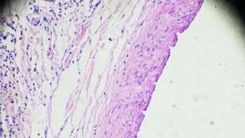
“With OCT you’re looking at a vertical section similar to histology (the study of the microscopic structure of tissues),” Orit Markowitz, M.D., director of Pigmented Lesions and Skin Cancer, at Mount Sinai Hospital, said in an interview with CURE. “You’re looking at a wider field of view and you’re also looking at, instead of one vertical section like you would with histology, a sort of flip book animation of vertical sections across the lesion. Although cellular clarity is not as good as a confocal microscope, depth is better.”
In addition, Markowitz noted that when OCT is used in combination with reflectance confocal microscopy (RCM) — another type of non-invasive imaging device that allows a dermatologist to see things in a cross-sectional view — it offers a three-dimensional view of a lesion that can’t be accessed even by cutting the skin, let alone with the naked eye.
“You’re able to not only make a diagnosis with RCM, but with OCT you can also also properly follow non-melanoma skin cancer lesions that you decide not to treat non-invasively,” she said. “Also, let’s say one small area of the lesion is of concern, then you can hone in on it with for the confocal microscope, and look at that small area to make a diagnosis.”
There are some limitations to non-invasive imaging, such as the time to scan and the possibility of misdiagnosing, if markings that don’t have clinical suspicion are scanned. Also, the OCT device is best used for nonmelanoma skin cancers.
However, non-invasive procedures help with better cosmetic outcomes, especially in areas that are cosmetically sensitive like the face, head and neck.
“When you cut skin that is scarred you have to go around the scar to see what is the extent of tumor involvement,” said Markowitz. “If I already have a scar from the biopsy, now the surgeon has to go around that scar. But, what if the lesion was smaller than the scar from the biopsy or what if the lesion was the same size?”
At this time, OCT is not covered by insurance, but Markowitz said the procedure would run a patient a couple hundred dollars out of pocket.
To prevent or catch skin cancer early, Markowitz advised that annual skin exams should start as soon as the average person is shifting from a pediatric doctor to an adult doctor. If they are at higher risk, such as having a primary relative with the disease or they have large atypical moles, then they should start getting screened earlier.
“A melanoma that is caught less than one millimeter thick, which is like the tip of a pencil compared to the tip of a crayon, now we are talking about more than a millimeter thick, can be the difference of life and death,” added Markowitz.
In addition to skin exams, people should wear sunblock daily, all year-round, avoid tanning beds and wear protective clothing to lower their risk of skin cancer. It is also wise to know the
“If they are coming in regularly for a skin exam, I tell my patients if you see something new, dark and concerning pre-ABCDE — you should probably get it looked at,” Markowitz said. “If you see something bleeding, crusting and not healing pre-ABCDE — I want you to come in and see me.”
Mount Sinai is offering free skin cancer screenings this month. For locations and times, click
Advertisement
Related Content
Advertisement


Advertisement
Advertisement
Trending on CURE
1
FDA Updates Safety Warnings for Common Chemotherapy Drugs
2
Real-World Data with Amtagvi Show High Response Rates in Advanced Melanoma
3
Chemo Brain is Real: My Experience with a Clinical Study and What It Can Teach Us
4
New Combination Treatment Approach Studied in Pancreatic Cancer
5