News|Videos|October 27, 2017
Follow-Up Care for Neuroendocrine Tumors
Advertisement
Episodes in this series
Transcript:
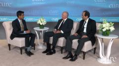
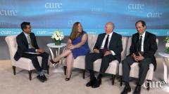
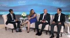
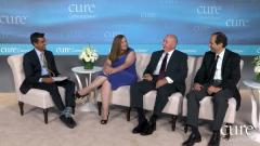
Shubham Pant, MD: Let’s move on to our next segment, which is really what we’ve talked about: taking ownership of your disease. Maryann, Larry, both of you took the disease with both hands and ran with it. So, how important—Maryann, I’ll come to you first—is it to take ownership of your disease, be an advocate for the disease, maybe join the Neuroendocrine Cancer Awareness Network?
Maryann Wahmann: Right. I think that, in the beginning, a lot of patients are scared, but I always tell the patients that they need to ask questions. The doctors might not know everything, try to get as much material as possible and bring the information to them. Each patient is different, each doctor is different. Find a doctor who will work with you closely, and open communication is the most important thing.
It wasn’t until after my diagnosis that I went back to my hematologist. I run fevers at night from the lupus. And I started running fevers during the day, or which I believed were fevers when I actually was having a flushing. Now had I mentioned that to the doctor when he would ask, “How are your fevers?” And I’d say, “I still had them.” Had I said to him, “Well, they’re starting to come during the day,” maybe I would have picked it up a little sooner. He said, “Why didn’t you tell me? I could have noticed.”
Shubham Pant, MD: Self-awareness is very important.
Maryann Wahmann: It’s very important. And a lot of patients think, “Well, I have this disease, I don’t have to worry about anything else.” Just as any other person in the world, you’re still prone to prostate cancer, breast cancer and everything else. Just because you have this cancer, it does not discriminate you from having any other cancer. And most important is that you have to be your own advocate. Make sure you go for your blood work, go for your CAT scans, go for your MIs, go for your Gallium scans. Make sure you stay on top of it. Don’t let it go.
I met a patient who had a Whipple procedure and her doctors told her she was cured, and I kept on saying, “You really need to be followed up.” And she’s like, “Nah, everybody said I’m cured.” She went and it turned out that she had another tumor, it was originally slow growing and it turned into a faster growing tumor. Had she not gone for her scans, she might have not been here. So, it’s important to stay on top of it, don’t wait five years to have the scans done. Go.
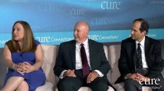
Shubham Pant, MD: What’s the regular follow-up for patients, Dr. Morse? Everybody’s different but do you have a path that you follow? I know every doctor is different in that, too.
Michael A. Morse, MD: The patients who have had primary surgeries and the cancer is thought to be gone, I agree, still need some follow-up; we would say early on. And there are some guidelines out there that can be used but it’s six-month intervals—CT scans or MRI scans, particularly of the area where they have the tumor and the liver. As time goes on, you can stretch it out to yearly. Because they’re so slow growing, some guidelines would suggest following people for 10 years, perhaps longer if you have reason to believe they could still relapse that far out. Somebody with known metastatic disease spread to the liver or elsewhere and with off-and-on therapy, if we’re actively treating, if the disease is changing, we may image it three to four months. If people are stable, it’s typically about 6-month intervals. And again, we prefer, because of their simplicity, CT scans or MRI.
You did bring up a new test, the Gallium-68 DOTATATE test. It’s important that people know about this test because it’s done as a CAT scan and as a PET scan together, but it’s not the typical PET scan that most people have heard of. And I think we’ve had scenarios where people came to see us and they said, “Well, I’ve had a PET scan,” and we’re trying to understand what PET scan did they have? They probably had the more typical one, which is called the FDG-PET scan. Those will only light up in the high-grade poorly differentiated tumors. They’re generally negative in the lower grade tumors.
The older test that had been done for lower-grade neuroendocrine tumors was called OctreoScan. The problem with OctreoScan is it has very poor resolution and you can miss lesions with it. Certain parts of the body, such as the liver and spleen, will light up on that test anyway and it’s a little bit difficult to differentiate tumor from background. Of course, it’s excreted into the urine so the kidneys and the bladder will have high uptake. With this newer type of PET scan, Gallium-68 DOTATATE, it’s very specific for neuroendocrine tumors and it’s very sensitive as well.
On the other hand, because it’s new, many insurance companies don’t seem to understand what it is, and there have been some challenges. And I think this is definitely where a person needs to be an advocate and if you get one denial, it’s try again. I had a recent scenario where we’re finding very often now that insurance companies are now hiring other companies to assess whether they should pay for the imaging. You and I spend hours a day dealing with this sort of thing. And it’s important to say, “This is new, it doesn’t go into the usual rubrics of the type of tests—the PET scans, the CTs—it’s different. And if they don’t pay for it today, if they get enough education, they’ll probably start paying for these because it’s a better test.”
And the hope is that in each person, their scenario is going to be different, but that we find a regimen that’s affordable, that’s easy for the person to comply with. It gives us the most information. Because if we can find it early recurring, as you pointed out, you can jump on it early and hopefully when it’s a smaller volume of disease.
Shubham Pant, MD: So, it’s care for individual patients. It’s not one-size-fits-all.
Transcript Edited for Clarity
Advertisement
Related Content


Advertisement
Advertisement
Trending on CURE
1
Balance Exercises for Patients With Cancer: How the Single-Leg Stand Builds Stability
2
Wildfire Smoke and Cancer Risk: What Patients Should Know
3
Clock Touches: An Advanced Balance Exercise for Patients with Cancer
4
Family History of Cancer Fuels Oncology Nurse's Passion for Patient Care
5