|Videos|March 29, 2019
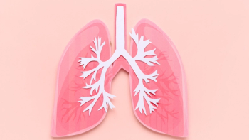
Role of Imaging in Diagnosing Lung Cancer
Advertisement
Episodes in this series


Transcript: Segment Description: The role of imaging modalities in confirming a diagnosis of lung cancer.
Edward Kim, M.D., FACP: We don’t have a lot of debates with chest MRIs because that is in itself a very inconvenient procedure. You’re in a machine for a long period of time. There is a role for MRI in certain situations, but a CT scan is still the best test for evaluation and diagnosis. And I tell people, the radiation exposure isn’t so bad because if you live in a high altitude or are on planes all the time, you’re having just as much exposure.
Philippa Cheetham, M.D.: Right. And yet even though you tell patients that, they are still very scared about radiation exposure. I’m sure you get asked a lot about the concerns about radiation from CT scans. And these patients are not just getting one CT of the chest, are they? They’re getting followed over many months. That’s not a concern?
Edward Kim, M.D., FACP: No, it’s all a risk/benefit. I think if we’re following active disease and treatments, it becomes less of a risk. If it’s long-term follow-up, then we start trying to think how we can space it out.
Philippa Cheetham, M.D.: I’m just talking about the CT scan to image the chest. Do patients need to have contrast, intravenous contrast to see lesions? Or is it better to avoid contrast?
Edward Kim, M.D., FACP: We prefer contrast because it will give us better imaging of specific structures, especially through the blood vessels, and there’s a lot of them, especially in the central part of the chest, or the mediastinum as we call it. We can do it without contrast if there is some kidney issue or if a patient’s older, but we prefer contrast.
Philippa Cheetham, M.D.: Now as a lung specialist I would think that if I looked at a CT chest of a nonsmoker, young, healthy person, and saw an abnormality, it would be much easier to identify an abnormal area that looked suspicious for a tumor than trying to look at a CT scan in somebody who’s got a lot of fibrosis or chronic obstructive airways disease or maybe signs of previous infections. How difficult is it for your radiologist to look through the jungle of chronic lung disease and identify suspicious lesions?
Edward Kim, M.D., FACP: I think if we have a high suspicion or see something abnormal, that certainly helps. From a pure screening standpoint, if someone had a lot of emphysema and lung changes, it’s very hard to detect. But they will learn. There have been lots of mammography publications over the years on the density of breast tissue and how to use techniques to sort out whether it’s calcifications or different techniques to assess dense areas. I think in lung cancer it will be the same thing, just as you say, as more screening occurs.
Philippa Cheetham, M.D.: Right. And we’ve already talked about the role of re-read on the pathology and how important it is that you get the biopsy done by the right person at the right time, and particularly for lesions that may be more challenging to biopsy. But even radiology, that is interpreted by a human being, and we know that the quality of machines differs, the strength of the imaging with MRI certainly, and that certain CT scans, the protocol that may be set up to image the chest, the software on the machine, and of course the human being who’s actually looking at those images. Do you see a huge variation in the quality of the report when you compare centers of excellence versus imaging in community hospitals?
Edward Kim, M.D., FACP: I think it is really important to have someone who does it as their day job to look at lung films. And they could be kidney films, or it could be brain films, but to have more subspecialty is a nice luxury. Now again, in some community hospitals you don’t have 30 radiologists who can divide their time equally to each of the different subspecialties. But it is certainly nice to have someone who’s used to doing it. There are different protocols, as you say, even the cut, sort of thickness, of CT scans varies.
Philippa Cheetham M.D.: Right.
Edward Kim, M.D., FACP: Between 1-1/2 mm to 5 mm. Gosh, I even saw a 7 mm the other day. I didn’t even know those machines still existed out there. But if you look hard enough, they probably do. And so I don’t think there’s an appreciation out there from the patient side that actually the type of machine, and not just the newest machine, but the type of machine, the type of technique, when you inject the contrast and then take the picture — all of that is an art form that is very similar to the technique of getting a biopsy. And then the read is the second part of it.
Philippa Cheetham, M.D.: The problem is as the patient, you don’t know what’s the quality of the machine or the software or even who’s looking at your imaging. And so many facilities are so great at marketing, we’ve got the latest, the greatest, the best machine. It’s very difficult I think for patients to know the quality of the imaging they’ve had. But one thing that always amazes me is when patients come for a second opinion. Often they arrive without any imaging, without any reports, with no pathology, and we’re expected to be the mind reader and the magician that offers a second opinion without any information. I think it’s so important to tell patients, if you’re going for a second opinion, take the copy of your report, get your images on a CD that you can take. Do you often open up images from outside facilities and review those?
Edward Kim, M.D., FACP: I completely agree. The patient, any time they have a medical record, and I admit I’m not probably the best at this either, needs to keep a copy for themselves. And it’s so important when you visit these other facilities, you want to maximize your time.
Philippa Cheetham, M.D.: Right, absolutely.
Edward Kim, M.D., FACP: You don’t want to go see someone for a second opinion only to hear them say, “I have no information in front of me, and can’t give you an opinion, and so you need to come back in a week after we get all the information, and talk about this again.” If you have the information there, I’ve done it many times, you put it in the machine, or you upload the images, you look at those aspects. We a lot of times want to get our own pathology reads before the visit so we have the pathology sent.
Philippa Cheetham, M.D.: That’s a good idea.
Edward Kim, M.D., FACP: I will say once even I had to walk across the street when I was in Houston to a different hospital facility because the pathology department there would not release the tissue. And I tell patients, “This is your tissue. It’s owned by you. Those scans, those are of you. You get to have those scans.” There seems to be this artificial barrier where patients feel like when it’s confined within a hospital wall somewhere that they can’t take it with them.
Philippa Cheetham, M.D.: They have no ownership.
Edward Kim, M.D., FACP: That’s right.
Philippa Cheetham, M.D.: Well, it’s a great point that you bring up, and I think it’s important that we use this opportunity to educate patients that it’s not just about getting a second opinion but providing the right information, particularly for a diagnosis like small cell lung cancer where timing is of the essence. And with a potentially aggressive disease you want to get on with making that diagnosis and start treatment as quickly as possible.
Transcript Edited for Clarity
Advertisement
Related Content


Advertisement
Advertisement
Trending on CURE
1
Balance Exercises for Patients With Cancer: How the Single-Leg Stand Builds Stability
2
Clock Touches: An Advanced Balance Exercise for Patients with Cancer
3
Wildfire Smoke and Cancer Risk: What Patients Should Know
4
Family History of Cancer Fuels Oncology Nurse's Passion for Patient Care
5