laBCC: Skin Biopsy
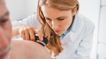
An expert discusses features of a skin lesion that would prompt a skin biopsy in the diagnosis of locally advanced basal cell carcinoma (laBCC).
Anna C. Pavlick, D.O.: Biopsies do not have to be done on every skin lesion that a patient has, thanks to the rigorous training of our dermatologists, who are very effective in identifying lesions that need to be biopsied. As people get older, they may also develop what are called skin tags. These are excessive tags of skin that occur as people get older; those are benign and don’t need to be biopsied. However, lesions that are new, that are bleeding and that just won’t heal—those are really the most common complaint for patients with basal cell. They will say, “I’ve got this little thing on my nose, and it just won’t heal. It scabs over, the scab falls off, it starts to bleed, it scabs again and then it just will not heal.” Those nonhealing lesions on people’s faces, heads, or necks are really a signal that says, “You might want to see your dermatologist about that.” Lesions that are scaly, skin lesions that are new or growing—sometimes these lesions can become ulcerated, meaning that they can bleed. They become like an open sore. They can look like a crater. They can get raised. They can get scaly. It takes the very skilled eye of our dermatologists to look at these lesions and say, “Hey, that needs to be biopsied because it’s looking abnormal, so we best take care of it while it is in its infancy because it makes management so much easier.”
There are several biopsies that a dermatologist can perform. The first and most common is called a shave biopsy, and a shave biopsy essentially occurs when a dermatologist will shave off the top part of a lesion and place it in a jar of formalin and send it to the pathologist. The pathologist will then look at it under the microscope and determine what that lesion is, what those cells are that they’re looking at. Are they cancerous? Are they not cancerous? The pathology report will also then tell us a) what type of cancer it is and b) how big it is—at least how big the specimen is or the cancer that they see on the cells that they have been analyzing, and sometimes they look at other features. Does it extend to the margins? Are there other adjacent microscopic tumors that may be near this primary tumor? We can really conclude a lot of information from the pathology report that helps us to best manage these skin cancers once we know what kind of skin cancer it is.
The other type of procedure that dermatologists can do to biopsy skin lesions is what’s called a punch biopsy, and a punch biopsy is when, as opposed to just shaving the top part of the skin lesion, we’ll punch down into the skin. It won’t get out the entire lesion. This certainly isn’t a curative procedure, but this will allow us to better examine how deeply this lesion goes down into the layers of the skin. Sometimes those are the recommended biopsies. Each dermatologist—based on the lesion that the patient has—is going to recommend a different type of a biopsy be performed. All the biopsies are sent to a dermatopathologist, a pathologist who specializes in reading skin types of pathology. Then we get back our report, essentially telling us what type of skin cancer this is and helping us determine how we best manage it for that patient.
Transcript edited for clarity.


