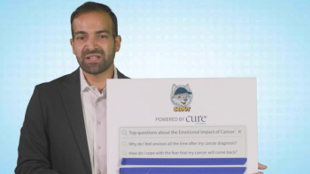
A psycho-oncologist answers 5 common questions about cancer-related anxiety, depression, fear of recurrence and emotional well-being.
Learn about lentigo maligna melanoma, from initial diagnosis and staging to the latest surgical treatments, systemic therapies and side effects.
Receiving a skin cancer diagnosis can feel overwhelming, but understanding the specific characteristics of your condition is the first step toward effective care. This educational overview of lentigo maligna melanoma, or LMM, is designed to help you understand your diagnosis, explore standard treatments and prepare for meaningful discussions with your oncology team.
Lentigo maligna melanoma is a distinct subtype of cutaneous malignant melanoma, which is a type of skin cancer that develops from pigment-producing cells called melanocytes. LMM typically arises on chronically sun-exposed skin, making the face, head and neck the most common locations. It is most frequently diagnosed in older adults, often peaking in a person's 60s and 70s.
This type of melanoma begins as a slow-growing, non-invasive patch known as lentigo maligna, or melanoma-in-situ. This means the cancer cells are confined strictly to the top layer of skin. Over time, sometimes spanning several years or even decades, it enters an invasive growth phase, penetrating into the deeper dermal layers of the skin. At that point, it is classified as lentigo maligna melanoma. Because it develops on severely sun-damaged skin, its borders are often ill-defined and can blend in with surrounding age spots or freckles, requiring careful mapping by specialists.
Diagnosing LMM requires a blend of visual mapping and microscopic evaluation. Because these lesions are common in highly visible areas like the face, doctors must balance diagnostic accuracy with cosmetic preservation.
During an initial evaluation, dermatologists utilize dermoscopy, a high-magnification light tool, to examine the unique pigment structures of the lesion. Advanced non-invasive tools, such as reflectance confocal microscopy, allow clinicians to view cellular layers down to 200 micrometers in depth to help map out exactly where the cancer ends and healthy skin begins.
A definitive diagnosis requires a tissue sample. While a complete removal is preferred, large lesions on the face may require a partial incisional biopsy or a specialized saucerization biopsy to sample the most suspicious areas along with adjacent normal skin.
Pathologists evaluate the tissue sample under a microscope using specialized stains called immunohistochemistry markers to confirm the presence of melanoma cells. LMM is staged using the American Joint Committee on Cancer system, which evaluates three main factors: The first factor is Breslow Thickness. This is the precise measurement of how deeply the melanoma has invaded the skin, measured to the nearest 0.1 millimeter. This is the single most critical factor in predicting prognosis and determining treatment width. The second factor is ulceration, which checks whether the top layer of skin over the tumor has broken down. This signifies a more aggressive disease state. The third factor is nodal involvement. If the melanoma is deeper, typically 1 millimeter or greater, or 0.75 millimeter with high-risk features, a sentinel lymph node biopsy may be recommended to check if microscopic cancer cells have spread to the nearest lymph nodes.
The primary goal of treating LMM is to achieve a complete cure while preserving as much healthy tissue and physical appearance as possible. Your treatment plan will depend directly on the depth and stage of the tumor.
Surgery remains the gold standard of care for localized LMM. Because LMM cells frequently extend invisibly past what the naked eye can see, standard safety margins are required.
A wide local excision involves the surgical removal of the tumor along with a predetermined margin of healthy surrounding tissue to ensure no cancer cells are left behind.
For LMM on the face or neck, doctors often utilize specialized margin-controlled techniques. In Mohs micrographic surgery or staged excisions, sometimes called "Slow Mohs," the surgeon removes the tissue layer by layer and examines 100 percent of the surgical margins under a microscope before closing the wound. This yields the lowest possible recurrence rates while conserving facial skin.
If a patient is not a good candidate for surgery due to age, health, or the size and location of the tumor, alternative options may be considered, though they carry a higher risk of the cancer returning.
Targeted radiation therapy can be utilized as a primary treatment for localized lesions that cannot be safely operated on.
In very specific, early cases or as an additional therapy, a topical cream called imiquimod may be prescribed to stimulate the body’s localized immune response to fight the abnormal cells.
If the pathology report indicates that the melanoma has spread beyond the local skin site to the lymph nodes or other parts of the body, systemic treatments are utilized. These include immunotherapies, which help your own immune system recognize and destroy cancer cells, or targeted therapies, which block specific genetic mutations if your tumor tests positive for alterations such as the BRAF gene.
Every cancer therapy carries potential side effects, which vary depending on the chosen treatment approach. Managing these effects is a collaborative effort between you and your care team.
Surgical excision can cause localized pain, swelling, bruising, scarring, risk of wound infection or localized nerve damage leading to temporary or permanent numbness.
Radiation therapy often leads to skin redness, irritation, dryness or peeling in the treated area, similar to a severe sunburn, along with localized hair loss and fatigue.
Topical imiquimod can trigger severe local skin reactions, including redness, blistering, crusting, itching and occasionally mild flu-like symptoms.
Systemic therapies can cause immune-related inflammation of healthy organs, such as colitis, pneumonitis, or skin rashes, as well as fatigue, joint pain or fever.
A diagnosis of lentigo maligna melanoma requires a highly personalized management strategy. Because this cancer frequently presents in cosmetically sensitive areas like the face and can possess invisible extensions beneath the skin, a multidisciplinary approach involving dermatologists, surgical oncologists and pathologists is vital to achieving the best outcome.
When you sit down with your oncologist, use this information to ask about the precise Breslow thickness of your lesion, whether margin-controlled surgeries like Mohs are appropriate for your specific location, and what to expect during the recovery process. Armed with the right questions, you and your doctor can chart a clear path forward toward a cure.
Editor's note: This article is for informational purposes only and is not a substitute for professional medical advice, as your own experience will be unique. Use this article to guide discussions with your oncologist. Content was generated with AI, reviewed by a human editor, but not independently verified by a medical professional.
For more news on cancer updates, research and education,