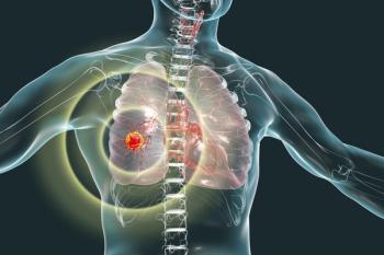
In patients with advanced ROS1-positive lung cancer who had not previously received a ROS1-targeted drug, Jideytro also shrank tumors that had spread to the brain in all 10 evaluable patients.
After receiving a stage 1 lung cancer diagnosis, Carolyn Rothfeld underwent robotic surgery to remove part of her lung and is back to her exercise routine nine months later.
She was almost done with her morning two-mile walk when a sudden jolt hit her upper right abdomen, right where her chest tube had once been. It felt like a static vibration, there and gone in a second, often triggered by something as small as a sneeze or a cough.
Carolyn Rothfeld called them "nerve jolts." Painless reminders, she said, of the surgery that saved her life nine-and-a-half months ago.
The journey started with a routine CT scan in March 2024, which incidentally picked up a small nodule in her lung. Dr. Keith Brenner, a pulmonologist at Hackensack University Medical Center, kept watch on it. A year later, another scan revealed the nodule had grown to 10 millimeters and appeared part solid, with the hazy, less-defined appearance known as a ground-glass opacity. Brenner told her he believed it was a stage 1A malignancy, then walked her to the office of Dr. Nabil Rizk, chief of thoracic surgery at John Theurer Cancer Center, who would lead her care from there.
"On the way down the hall, all of a sudden, I started to feel freezing cold," Rothfeld said. "My body was suddenly reacting to the news."
Brenner reassured her that everything was going to be alright. The surgery she'd need would be a robotic procedure — minimally invasive, designed to remove as little of her healthy lung as possible.
Within four weeks, Rizk performed the robotic video-assisted thoracoscopic surgery (VATS) segmentectomy to remove the cancerous portion of Rothfeld's lung. Biopsies confirmed that the adenocarcinoma was contained within the nodule and had not spread.
Dr. Geoffrey Pelz, a thoracic surgeon on Rizk's team, explained there are three general ways to biopsy a lung mass.
The first is a CT-guided biopsy, where the area is numbed from the outside, and a needle is guided in — useful for larger masses near the edge of the lung.
The second approach works from the inside of the airways, using a technology called ion robotic bronchoscopy. A CT scan is fed into a computer, which builds a 3D reconstruction of the lung and its airways.
"It's kind of like a GPS for your car," Pelz said. "I can say that this is the target I want to go to. It'll draw me a little roadmap, and then we can follow that in the operating room and reach any part of the lung from the inside."
The third option is to go straight to surgery — sometimes a limited wedge resection or another lung-sparing procedure, depending on the patient.
"I can give my opinion as to what I think is best for that patient, but ultimately it's up to the patient and family to decide," said Pelz.
Rizk explained that most lung surgeries today are done robotically, in a minimally invasive fashion — a shift that has dramatically improved healing time and recovery. In Rothfeld's case, the lesion sat too deep for a simple wedge resection, so her team performed a segmentectomy instead, sparing two-thirds of that part of her lung while still removing the cancer.
The decision about how much lung tissue to remove depends on the tumor's location and size, Rizk said. Smaller masses near the edge of the lung can often be removed with a localized resection, along with a sample of nearby lymph nodes to check for spread. Larger tumors, those located deeper in the lung, or those that span multiple segments sometimes require a lobectomy — or, in rarer cases, removal of an entire lung.
Rizk's team uses the ion bronchoscopy technology to inject a dye near the tumor before surgery. This helps locate tumors that are hard to see or feel. The process adds about five minutes to the procedure to give surgeons a visual guide once they're operating with the robot, helping ensure a clean margin.
"That's a technology we utilize fairly frequently in the operating room," Rizk said, "to be able to intervene on lung cancers earlier.”
Rizk said one of the biggest barriers to getting patients into treatment is fear left over from an earlier era of lung surgery. A thoracotomy used to include a large incision, divided muscle, ribs spread apart, followed by a long recovery.
"A lot of people may have known somebody who had surgery 15, 20 years ago," he said. "They think, oh, they're going to be in such excruciating pain for the rest of their lives."
Most patients stay in the hospital only overnight, he said. Once the lung has healed enough the next day, the chest tube comes out and the patient goes home. Most people return to normal activities within about two weeks, though full nerve healing can take two to three months.
Nine-and-a-half months later, Rothfeld is back at the gym and walking two miles almost daily. Her nerves are healing, and her mobility is returning.
"They're not painful. They're just reminders, and I know that's going to eventually subside completely," she said.
She said she never once regretted going through with the surgery. If anything, it gave her more time with her family — the people who showed up for her amid their own busy lives.
"Whatever your personality is, introvert, extrovert, whatever, the most important thing is not to isolate yourself," Rothfeld said. "You don't have to go through it alone."
For more news on cancer updates, research and education,